An Atomic Force Microscope, often known as an AFM, is a highly advanced tool that gives researchers the ability to view surfaces on a nanoscale scale. In contrast to conventional optical microscopes, which are constrained by the diffraction of light, atomic force microscopes (AFMs) are able to attain resolutions that are as low as fractions of a nanometer, which makes it possible to visually observe individual atoms and molecules.
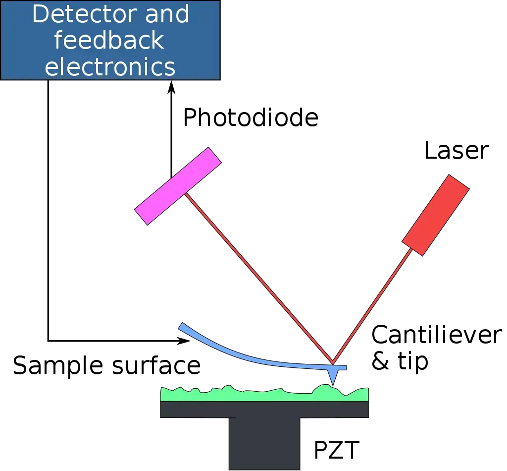
Cantilever is the name of the sharp probe that is used in the operation of an atomic force microscope (AFM), which is used to scan the surface of a material. During the process of moving over the surface, the probe is subjected to forces that are caused by the topography of the sample, which results in the cantilever deflecting. A laser beam that is reflected off of the cantilever and onto a position-sensitive photodetector is what is used to detect these deflections. The atomic force microscope (AFM) is able to generate a comprehensive topographic map of the surface by utilizing a feedback loop to ensure that a consistent force is maintained between the probe and the sample.
AFMs are extremely flexible instruments that are utilized in a wide range of scientific fields. Within the field of biology, they have been utilized for the purpose of analyzing protein structures, imaging biological membranes, and studying the mechanical characteristics of tissues and cells within the body.
At the nanoscale, atomic force microscopes (AFMs) are applied in the field of materials science to investigate surface roughness, texture, and other morphological characteristics.
Because it can function in a variety of conditions, including air, liquid, and vacuum, atomic force microscopy (AFM) is ideal for a broad variety of samples, ranging from hard materials like metals to soft biological tissues. This is one of the key advantages of AFM. Furthermore, atomic force microscopes do not need any particular sample preparation, such as staining or coating, which allows the natural condition of the material to be preserved for imaging purposes.
Since its inception in 1985 by scientists working for IBM, the atomic force microscope (AFM) has evolved into a vital instrument in the area of nanotechnology and other related sciences, offering insights into the nanoscale world that are unmatched.
Principle of Atomic Force Microscope
The Atomic Force Microscope operates on the basis of sensing intermolecular forces and detecting atoms utilizing probed specimen surfaces at the nanoscale. Its operation is facilitated by three primary working principles: surface sensing, detection, and imaging.
The Atomic Force Microscope (AFM) uses a cantilever to do surface sensing. The cantilever has a sharp tip that scans over the sample surface by creating an attractive force between the surface and the tip as it moves closer to it. When it comes into close contact with the surface of the sample, a repulsive force gradually takes control, causing the cantilever to move away from the surface.
As the cantilever deflects away from the sample surface, the direction of reflection of the beam changes, and a laser beam detects the aversion by reflecting off a beam from the cantilever’s flat surface. It captures these changes in deflection and direction of the reflected beam using a positive-sensitive photodiode (PSPD- a component based on silicon PIN diode technology that measures the position of the integral focus of an incoming light signal).
The Atomic Force Microscope (AFM) captures images of a sample’s surface topography by scanning the cantilever across a portion of interest. The Positive-sensitive photo-diode (PSDP) monitors the deflection of the beam, which is determined by how elevated or low the surface of the sample is. The microscope contains a feedback loop that adjusts the length of the cantilever tip slightly above the sample surface; hence, the laser location is maintained, resulting in an accurate imaging map of the image’s surface.
Parts of Atomic Force Microscope
- Cantilever—A thin, flexible beam with a sharp point. When the tip touches the sample, it bends to measure forces.
- Tip- The cantilever’s sharp probe examines the surface. Silicon or silicon nitride is used for durability and accuracy.
- Laser System—A laser beam targets the cantilever’s back. To track cantilever motion, the laser reflects onto a photodetector.
- photodetector — Converts laser deflection from cantilever bending into an electrical signal. This signal matches surface properties.
- Piezoelectric scanner—For nanometer-level precision, the scanner moves the sample or tip in x, y, and z directions.
- Feedback System- controls tip-sample distance. Imaging is done at constant force or height to avoid harm.
- Control electronics – The scanner, photodetector, and feedback mechanism are controlled by control electronics. This keeps the system running smoothly.
- Computer system—Converts instrument signals into pictures. It aids data storage and analysis.
- Sample holder—keeps sample stationary during scanning. It depends on sample size and kind.
- Vibration isolation technology — Prevents extraneous vibrations from disrupting images, maintaining precision.
- Optional accessories –With temperature controls or liquid cells, the AFM may be customized for studies.
Operating Procedure of Atomic Force Microscope (AFM)
Using an Atomic Force Microscope (AFM) requires numerous painstaking processes to guarantee good imaging and data capture. Here’s the overall procedure:
- System initialization:
- Power On: Turn on the AFM system, which includes the computer, controller, and vibration isolation unit.
- Launch the AFM control software to interact with the instrument.
- Cantilever and Tip Preparation:
- Select the proper cantilever and tip for the sample and imaging mode.
- Installation: Carefully attach the cantilever to the holder, ensuring it is secure and undamaged.
- Laser Alignment:
- Positioning: Focus the laser beam on the rear of the cantilever.
- Optimisation: Adjust the laser location to produce the best signal on the photodetector, usually aiming for a voltage response between 4 and 5 V.
- Sample Placement:
- Mounting: Place the sample on the stage, ensuring that it is clean and securely fastened to avoid movement during scanning.
- Focus Adjustment: Use the optical microscope to bring the sample surface into good view.
- Engaging The Tip:
- Approach: Using the software’s approach feature, gradually move the tip closer to the sample surface while watching for any abrupt contact that might harm the tip or sample.
- Scanning Parameter Setup:
- Mode Selection: Depending on the sample and analysis needs, select the appropriate imaging mode (contact, tapping, or non-contact).
Parameter Configuration: Set scanning parameters including scan size, pace, and feedback controls to improve image quality.
- Mode Selection: Depending on the sample and analysis needs, select the appropriate imaging mode (contact, tapping, or non-contact).
- Image Acquisition:
- Scanning: Start the scan and let the AFM to collect data over the specified region.
- Monitoring: Observe the scanning process in real time and modify settings as needed to improve image quality.
- Data Analysis and Storage:
- Processing: Use the program to process the collected data, producing visual and quantitative results.
Saving: Ensure that the data is properly stored for future analysis and documentation.
- Processing: Use the program to process the collected data, producing visual and quantitative results.
- System Shutdown:
- withdraw Tip: To prevent damage, safely withdraw the tip from the sample.
- Power off the AFM system and related components according to the manufacturer’s instructions.
Applications of Atomic Force Microscope
- Surface Characterization—AFM allows nanometer-scale surface topography imaging and analysis. This is needed to analyze surface roughness, texture, and morphology in materials research.
- Biological Research – AFM examines everything from large tissues to individual cells and proteins. Biological samples can be studied without staining, keeping their natural condition.
- Nanomechanical Measurements– Nanoscale mechanical qualities including elasticity, stiffness, and adhesion are measured using AFM. For polymer, thin film, and nanostructured material mechanical behavior evaluation, this is essential.
- Force Spectroscopy-distance to examine tip-sample interactions. Nanoscale contacts, atomic bonding, Van der Waals forces, and single-molecule stretching and rupture forces are measured using this method.
- Nanolithography—AFM manipulates materials at the nanoscale to create nanometer-sized shapes and patterns for electronics and photonics.
- Material Science—AFM examines polymers, thin films, coatings, piezoelectrics, ferroelectrics, graphene, and other 2D materials, revealing their surface characteristics and behaviors.
- Cancer Research — AFM can help distinguish malignant from normal cells by providing high-resolution ultrastructure and mechanical characteristics of tumor cells.
- Chemical Analysis — Working with other methods, AFM can map surfaces’ compositions to identify materials and chemical characteristics at the nanoscale.
- Environmental Science—AFM studies aerosols and nanoparticles to determine their morphology and mechanical characteristics, which helps assess their health and environmental impacts.
- AFM is used in the semiconductor industry to inspect and characterize wafer surfaces, detect faults, and ensure microfabrication quality.
Advantages of Atomic Force Microscope
- AFM can image surfaces at the atomic level, exceeding several conventional microscopy methods.
- Flexible Sample Analysis — It can evaluate polymers, ceramics, composites, glass, and biological samples regardless of electrical conductivity.
- Unlike electron microscopes, AFM requires little or no sample preparation, keeping the sample’s natural condition and saving time.
- AFM works in air, vacuum, and liquid settings, making it suited for examining biological samples in their natural state.
- Real 3D surface profiles give precise surface topography information needed for material characterisation.
- AFM measures nanoscale mechanical characteristics including adhesion, hardness, and friction to understand material behaviour.
- Using non-destructive testing, fragile samples may be analyzed without harm.
- Integrates with Other Techniques — AFM may be used with optical microscopy and spectroscopy, broadening its scientific applications.
Disadvantages of Atomic Force Microscope
- Limited Scan Size – The limited scan size of the AFM allows imaging of areas no larger than about 150×150 micrometers, and its maximum height range is limited to between 10 and 20 micrometers, which severely limits its ability to analyze larger samples.
- Slow Scanning Speed –Additionally, the scanning speed is relatively slow, often taking several minutes for each image, a factor that can lead to thermal drift and degrade measurement accuracy.
- Tip-Related Artifacts – Image quality depends very much on the geometry of the probe, and any damage or contamination at the tip will introduce artifacts into the data and compromise data integrity.
- Surface Interaction Limitations – AFM may not accurately measure steep walls or overhangs because of physical interaction between the tip and sample surface.
- Potential Sample Damage – Due to the contact modes of AFM, it may cause surface damage to soft or fragile samples, which can limit its applicability to certain materials.
- Environmental Sensitivity – AFM measurements are sensitive to environmental factors such as vibrations, temperature fluctuations, and so on, requiring controlled conditions for best operation.
- Complex Sample Preparation – Although much less sample preparation is required by AFM compared to electron microscopy, certain samples may still need to be prepared in a special way for the outcome to be valid.
- Limited Depth of Field – AFM has a limited depth of field compared to techniques such as Scanning Electron Microscopy that can capture wider areas with great depth.
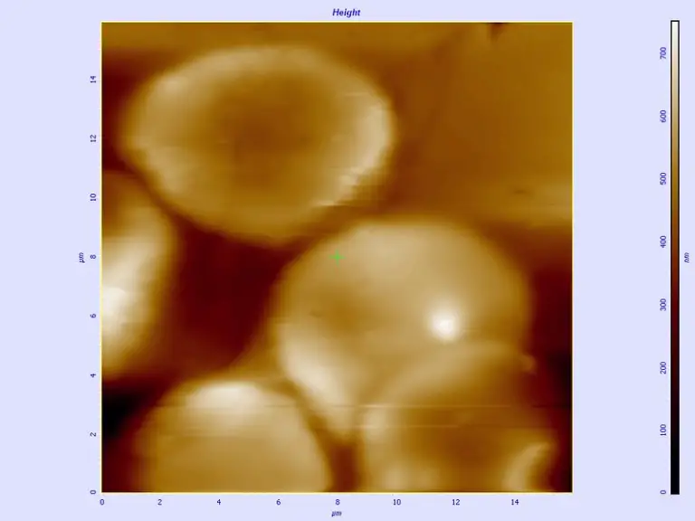
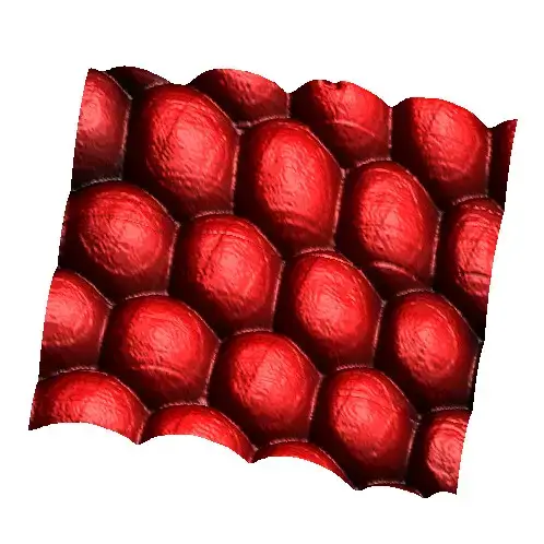
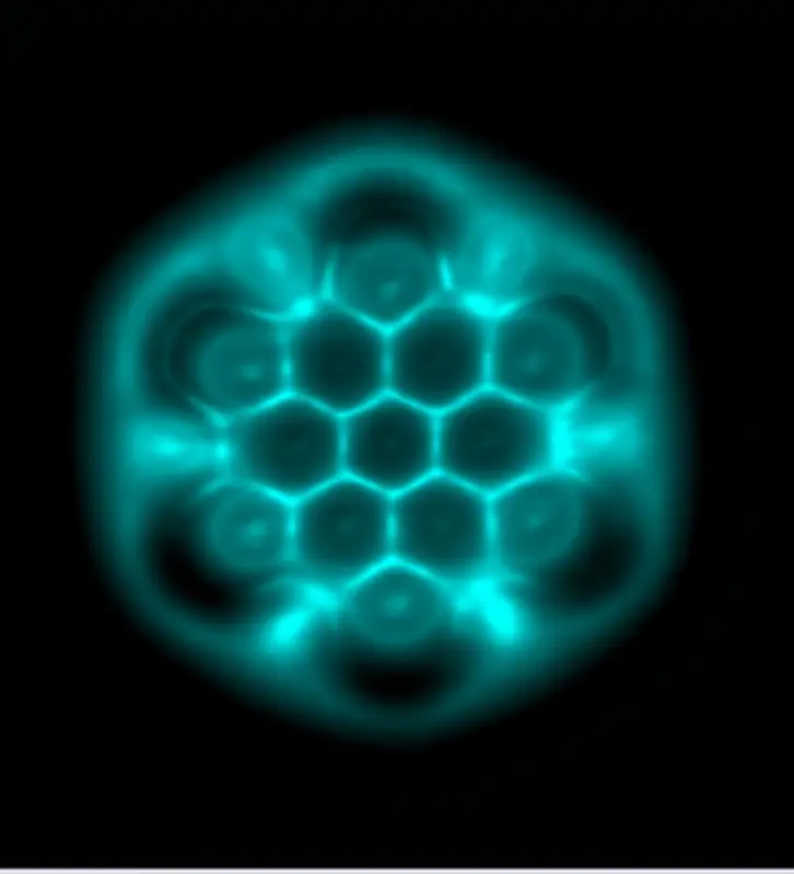
Atomic force microscope pictures/atomic force microscope images
- Binnig, G.; Quate, C. F.; Gerber, Ch. (1986). “Atomic Force Microscope”. Physical Review Letters. 56 (9): 930–933.
- Cappella, B; Dietler, G (1999). “Force-distance curves by atomic force microscopy”. Surface Science Reports. 34 (1–3): 1–104.
- Zhong, Q; Inniss, D; Kjoller, K; Elings, V (1993). “Fractured polymer/silica fiber surface studied by tapping mode atomic force microscopy”. Surface Science Letters. 290 (1): L688.
- Radmacher, M. (1997). “Measuring the elastic properties of biological samples with the AFM”. IEEE Eng Med Biol Mag. 16 (2): 47–57.
- Galvanetto, Nicola (2018). “Single-cell unroofing: probing topology and nanomechanics of native membranes”. Biochimica et Biophysica Acta (BBA) – Biomembranes. 1860 (12): 2532–2538.
- https://www.first-sensor.com/en/products/optical-sensors/detectors/position-sensitive-diodes-psd/
- https://www.researchgate.net/publication/256195163_The_Atomic_Force_Microscope
- https://www.sciencedirect.com/topics/nursing-and-health-professions/scanning-probe-microscope
- https://amedleyofpotpourri.blogspot.com/2018/09/atomic-force-microscopy.html
- http://nanoscience.gatech.edu/zlwang/research/afm.html