A digital microscope is a brilliant optical innovation, fundamentally distinguished from a normal microscope simply because it does not employ any eyepiece. The digital microscope uses a digital camera as its image detector and projects the images onto a computer monitor or screen. The entire process eliminates human interaction with the optical system; all the processes of viewing images are done electrically.
While a traditional microscope demands an external light source that would have to traverse through the eyepiece, a digital microscope usually contains an LED system itself for light. The very format of the digital microscope offers certain inbuilt advantages. For example, while several digital microscopes are USB digital microscopes, they still harbor advanced features like macro lenses and webcams. Higher models have specific illuminations such as Kohler and phase-contrast systems that improve the resolution and contrast of images to a considerable extent. They are primarily utilized in industries and by professionals.
The first digital microscope was invented in 1986 by Hirox Co. LTD, which was located in Tokyo, Japan. It consisted of a control box attached to the camera along with a lens that marked the beginning of computerized microscopy. Digital microscopes were to undergo great transformations with time. In 2005, a newer version was developed with an inbuilt monitor and computer that did away with the use of an external device. The new design made the device even more compact and easier to use. By 2015, digital microscopes had evolved into devices with USB connectivity to external computers, which increased processing speed but reduced the number of cumbersome cables.
Digital microscopes are equipped with advanced software intended to process and enhance images. It includes functions such as adjusting brightness, contrast, scaling, and cropping to focus on specific areas of interest. These features are quite essential, making digital microscopes very useful tools in educational and professional settings where high-quality, detailed imaging is of utmost importance.
Principle of a Digital Microscope
Digital Microscopes have hardware and software tools that enable the function of focusing and extracting an image from a specimen. The specimen’s image may be captured with the installation of software; whatever can be captured from the specimen appears on the computer monitor screen. These captured images might appear as static pictures in some cases, while others appear like motion videos. They can also be recorded, edited, cropped, labeled, and saved. The software can also be used for measuring the image sizes, magnifying, and modification the image in several ways.
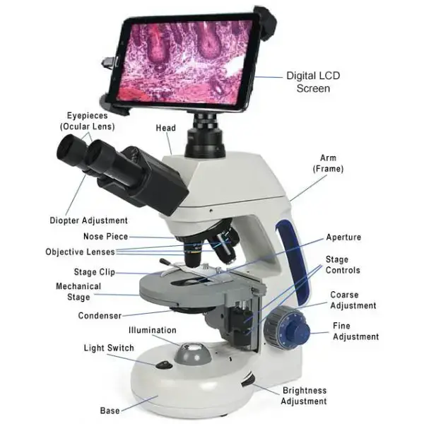
Parts of a Digital Microscope
- Hardware Components- Its comprised of the analogue section which includes a light source, traditional microscope structure and cam that substitutes for conventional eyepiece to display specimen images on computer screens.
- Software section– The programme includes organised units such as viewer component, brightness modifier, image contrast section and histogram equalisation capability to process visuals based on user preferences.
- Image Processing capability – Like most microscopes, it employs grayscale transformation equation (Jo = Ji + C) where Jo represents output visual, Ji shows input image whilst C indicates brightness adjustment constant.
- Viewer unit –This component continuously displays specimens and captures images from scope which are then stored and processed according to operator requirements.
- Brightness control– The unit enhances image illumination depending on light focused on specimen, it controls pixel intensity through positive or negative C values.
- Image scaling capability – They process and modify specimen dimensions whilst maintaining structural integrity of the visual data.
- Cropping function– Its designed to remove unwanted sections from captured images whilst preserving essential specimen details for analysis.
- Processing software– Contains specialised drivers and image manipulation programmes that work together to create high-quality specimen visuals.
- Display mechanism– The cam focuses specimen image which then appears on computer monitor where its stored and can undergo further processing depending on requirements.
- Light source placement –The illumination component position varies based on microscope type, with some having it beneath stage whilst others place it above.
- Camera integration– Most digital scopes utilise removable cams connected via C-mount adapter, though some models feature permanent USB connections.
- Monitor interface– These instruments connect to computer displays which serve as both viewing and control mechanisms for specimen analysis.
- Image storage– Its capable of preserving vast quantities of specimen data through sophisticated recording and storage mechanisms.
- Processing units– The software contains multiple specialised sections that work together to enhance and modify captured specimen images.
Types of Digital Microscopes
- Biological Digital microscopes – Its contains high magnification capabilities with light source beneath mechanical stage, providing 4x-100x objective with halogen or LED illumination for detailed specimen analysis.
- fluorescence Digital Microscopes– Like most microscopes, these optical devices utilise fluorescence and phosphorescence light sources to generate specimen images.
- inverted digital scope– The instrument contains light source and condenser positioned atop stage whilst objectives remain below for specialised examination through trinocular design.
- Metallurgical Digital Microscopes – Its specifically designed to examine metallic surfaces, wire circuits and opaque specimens through sophisticated illumination techniques.
- phase digital microscopes– They observe unstained specimens in both living and non-living states through inverted or upright configurations.
- Stereo Digital scope– The device reflects light from specimens whilst examining electric components, artefacts, Plants, circuitry and Art through sophisticated optical systems.
- polarising Digital Microscopes– Contains specialised video cam combined with high magnification lens and multi-ultra-bright LEDs that evaluate 3D structures of anisotropic specimens through polarised light waves.
- Digital USB scope– Its permanently connected to computers via USB connector, unlike traditional digital microscopes that utilise removable C-mount adapter cams.
- handheld Digital Microscopes – They integrate modern microscope systems for surface inspection and forensic analysis through portable design.
- portable digital scope– These compact wireless instruments examine hard-to-reach surfaces, making them ideal for medical examinations, field inspections and dermatological studies through sophisticated imaging capabilities.
Uses of Digital Microscopes
Here are various applications of Digital Microscopes in different sectors:
- Cytological studies– Scientists utilise these devices to examine cell structures and patterns in great detail, which helps them understand cellular behaviour and identify abnormalities.
- Brewing Industries – These microscopes analyse yeast cells and monitor fermentation processes, ensuring proper quality control during beer and wine production.
- Water and Waste Treatment– Digital scopes help technicians inspect microorganisms and contaminants in water samples, which is crucial for maintaining water quality standards and treatment efficiency.
- Forensics studies – Like most microscopes they assist investigators to examine trace evidence, hair samples, and fibres at crime scenes, whilst providing detailed digital documentation of findings.
- Dental Studies– These instruments enable dentists to examine tooth structure, identify cavities, and perform precise procedures. They are particularly useful for root canal treatments and microsurgery.
- Fetal and embryonic Transplant studies – The stereoscopic microscope helps scientists observe developmental stages and analyse tissue samples for research purposes.
- Microsurgical procedures – Doctors use digital scopes to perform delicate operations with high precision, whilst the stereo microscope provides enhanced visualization of the surgical site.
- Transplantation procedures– These devices aid surgeons in examining tissue compatibility and monitoring post-transplant healing processes, which requires detailed observation at the cellular level.
Advantages of Digital Microscopes
- High-resolution magnification– These microscopes produce crystal-clear images measured in pixels, which enables researchers and technicians to examine specimens with exceptional detail and clarity.
- Utilting capabilities – It provides both 2D and 3D measurements of specimens whilst allowing users to view objects from multiple angles, which proves invaluable for comprehensive analysis.
- Data Storage– Like most digital devices, they store massive amounts of visual information through imaging and recording of both static and dynamic specimens, making it easier to document and track changes over time.
- One-stop operation machines – These microscopes visualise specimens and display results directly on computer monitors, which streamlines the entire examination process whilst reducing the need for additional equipment.
- Cost-effective– Some digital microscopes are available at reasonable prices, making them accessible to smaller laboratories, educational institutions and individual researchers.
Limitations of Digital Microscopes
- Digital resolution- They have limited resolution compared to traditional optical microscopes, which can affect image quality when examining extremely small specimens.
- Cost of high-end models– Like most sophisticated equipment, advanced digital microscopes with premium features can be quite expensive, making them inaccessible for smaller organisations.
- Software compatibility issues – The scope often requires specific software that might not work with all operating systems, whilst updates can cause operational problems.
- maintenance requirements– These instruments need regular calibration and maintenance to ensure accurate measurements, which can be time-consuming and costly.
- Image processing limitations – The stereoscopic microscope sometimes produces digital artifacts or noise in images, particularly in low-light conditions.
- Storage constraints– It requires substantial digital storage space for high-resolution images and videos, which can strain computing resources.
- Digital eye strain- Users may experience visual fatigue from prolonged viewing of digital displays, unlike traditional microscopes that utilise direct optical viewing.
- Field of view restrictions – They provide a more limited field of view compared to conventional microscopes, which can make it challenging to examine larger specimens.