An inverted microscope is a special type of microscope. It doesn’t resemble other regular microscopes, as the light source and condenser point upward to shine directly downwards onto the specimen stage. The objective lenses, along with the turret, face up from below the stage. In other regular upright microscopes, the stages have objectives facing up while the light source is pointing down. It was invented by J. Lawrence Smith in 1850 at the Medical College of Louisiana, which is now Tulane University. In general, an inverted microscope has a fixed stage to hold bigger sample containers, like Petri dishes or tissue culture flasks, so it can better observe living cells or organisms under more ideal conditions.
Focus adjustment is achieved by moving the objective lenses along a vertical axis to bring them closer to or farther from the specimen, utilizing dual concentric knobs for coarse and fine adjustments. A rotating turret, or nosepiece, holds multiple objective lenses of varying magnifications, allowing for versatility in viewing. In addition, cameras for photomicrography and movie-making, fluorescence illumination, and confocal scanning units can be added to inverted microscopes to improve their operation. There are several advantages of inverted microscopes. The primary advantage of inverted microscopes is that a person can observe living cells or organisms placed at the bottom of large containers, like tissue culture flasks, without having to move the sample onto a glass slide. This feature is very useful in cell biology and microbiology, where the maintenance of the original environment of the specimen is highly critical.
Inverted microscopes are also used in micromanipulation procedures, providing enough space above the specimen for tools and equipment. In metallurgy, they help to observe polished specimens mounted on the stage, which can be observed from below through specific lenses.
Principle of the Inverted Microscope
Like a normal light microscope, an inverted microscope works by using the same basic ideas. In order to generate an image that can be viewed by the objective lenses, they employ light rays to focus on a specimen. However, in the inverted microscope, the light source and condenser are located on top of the stage, looking downwards. The primary function of the condenser lens located above the specimen stage is to focus the light on the specimen. The sample is put on a big stage that can hold it. The objectives, which are placed below the stage and pointed upwards, collect light from the condenser, magnifying the image before sending it to the ocular lens. The ocular lens reflects light through a mirror. With the help of an ibidi Polymer Coverslip and an ibidi Glass coverslip, the cells can be seen and studied through the bottom part of the cell culture device, which has total optical points.
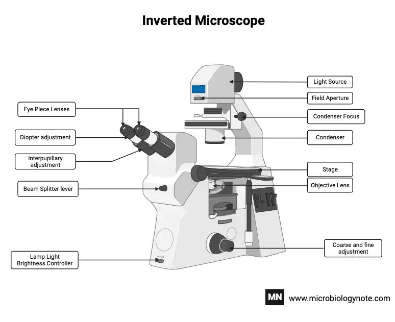
Parts of the Inverted Microscope
The main parts of an inverted microscope include:
- Light Source: This is the source of light to the specimen. In an inverted microscope, the light source is above the stage and shines down on the specimen.
- Condenser Lens: This focuses light on the specimen. It is located above the stage and collects light from the source to shine on the specimen, thus making it brighter and clearer.
- Stage: Stabilizes the specimen. It is generally bigger and remains stationary, designed to hold several sample containers such as Petri dishes or culture flasks.
- Objective Lenses: Magnify the specimen. These are positioned underneath the stage, which they face upwards, hence capturing and enlarging the view of the specimen. A few objectives vary in magnification and are fixed to a rotating nosepiece.
- Nosepiece (Turret): It carries and rotates the objective lenses. Different magnifications can be selected by rotating the desired objective to its position.
- Focus Adjustment Knobs: Fine focusing is done using the knobs. Dual concentric knobs, which have coarse and fine adjustments, move the objective lenses vertically for getting the exact focus on the specimen.
- Eyepieces (Ocular Lenses): These are used to observe the magnified image. They are placed at the top of the microscope and further magnify the image formed by the objective lenses.
- Illumination Control: It controls the intensity of light. Diaphragms and filters control the amount and quality of light reaching the specimen to enhance contrast and clarity of the image.
Uses of the Inverted Microscope
- Living Cell Imaging: Inverted microscopes are suitable for natural cell observation. They let scientists study cells in growth dishes without touching them, which makes it easier to see how cells are working in real time.
- Industrial Applications: To analyze huge or heavy samples that upright microscopes cannot handle, inverted microscopes are utilized in industrial settings. They provide below-the-surface inspection of metallurgical samples, electronic components, and other materials, allowing sample examination flexibility.
- Micromanipulation: Inverted microscopes are well-suited for micromanipulation techniques due to their ample space above the stage. This includes techniques like in-vitro fertilization (IVF), which need to carefully change cells or embryos.
- Time-Lapse Imaging: Inverted microscopes enable researchers to observe dynamic processes in living cells over extended periods, thereby facilitating time-lapse studies. This is key to understanding cell development, division, and stimulus response.
- Material Science: Inverted microscopes inspect surfaces, detect faults, and investigate metal and alloy microstructures. Their configuration enables the investigation of samples that are difficult to see with ordinary upright microscopes.
Advantages of Inverted Microscope
- Living Cell Observation: Inverted microscopes let researchers see living cells without pretreatment. This is useful in cell culture experiments when cells stick to culture containers.
- Larger Samples: Inverted microscopes can examine larger and heavier samples than upright microscopes. Industrial applications with tall or bulky samples benefit from this.
- Enhanced Sterility: Objective lenses are below the stage, so they don’t touch the sample. This arrangement prevents contamination during live cell imaging by maintaining sterility.
- Easy Sample Access: The inverted structure allows access to the sample from above, making medium exchange and micropipetting easier.
- Avoiding Objective Damage: By placing the objectives underneath the level, sample manipulation can be done without accidently destroying them.
- Time Efficiency: Inverted microscopes provide faster sample changes and adjustments because the specimen does not need to be repositioned. This efficiency benefits research and industry.
Limitations of Inverted Microscope
- Limited Magnification – An inverted microscope may not be able to obtain the extraordinarily high magnifications that specialized upright or electron microscopes can provide, yet being appropriate for a wide range of biological and industrial applications.
- Limited Field of View- They frequently have a lower field of view than upright microscopes, which might make it difficult to study larger objects.
- Sample Size – These microscopes are for thin, flat, or liquid samples. Bulky specimens may not fit or block objective lens focus.
- Restrictions on technique flexibility- Some sophisticated imaging techniques, such as polarized light microscopy, might not work as well with an inverted microscope’s architecture.
- Price— Inverted microscopes cost more than upright ones due to their unique shape.
- Inconvenience in Specific Uses – When working with opaque samples, the illumination may not be as effective because it originates from below the stage.
- Maintenance—The complicated optical structure, including lenses below the stage, makes cleaning and maintenance harder.
- Ergonomic Challenges- Despite being designed for ease of use, their design may nonetheless cause uncomfortable positions after extended use.
- Not beginner-friendly– Upright microscopes are frequently easier to use, making them ideal for instructional and beginning applications.
Inverted Fluorescent Microscope worksheet
- Wei, H., Chen, J., Wang, S., Fu, F., Zhu, X., Wu, C., Liu, Z., Zhong, G., & Lin, J. (2019). A Nanodrug Consisting Of Doxorubicin And Exosome Derived From Mesenchymal Stem Cells For Osteosarcoma Treatment In Vitro. International journal of nanomedicine, 14, 8603–8610. https://doi.org/10.2147/IJN.S218988
- Sun, Y., Wang, D., Ma, Y., Guan, H., Liang, H., & Zhao, X. (2019). Elucidating Escherichia Coli O157:H7 Colonization and Internalization in Cucumbers Using an Inverted Fluorescence Microscope and Hyperspectral Microscopy. Microorganisms, 7(11), 499.
- https://doi.org/10.3390/microorganisms7110499https://conductscience.com/lab/inverted-fluorescent-microscope/