What is a Dissecting microscope (Stereo microscope)?
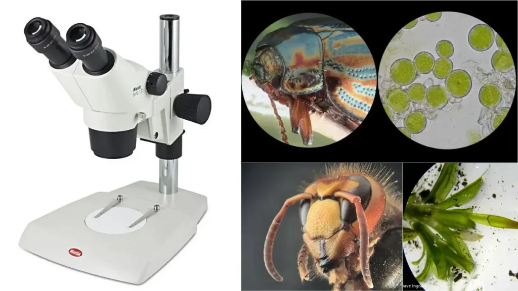
Dissecting microscope (stereo microscope), It’s typically used to look at small things or for dissections or soldering, activities that need close-up viewing.
Dissecting microscopes generally offer low magnification levels (approximately 4x to 50x) and have a relatively large working distance so that the object being viewed may be manipulated by hand using forceps or tweezers. Mainly biology, entomology, geology, and engineering use them.
A stereo microscope is composed of two eyepieces, each having its objective lens. These are affixed to a stand that is adjustable in both height and angle. The object that is being viewed lies on a stage that can be raised and lowered or moved around as desired to focus the image. Some dissecting scopes even come with a lamp or L.E.D.
This in turn led to the discovery of the dissecting microscope, first made in the 17th century with Anton Van Leeuwenhoek’s microscope of a simple single lens, a copia of which is on display in the R.O.M. But today’s stereo microscope did not arrive until the 19th century.
At the dawn of the 19th century, various scientists and inventors began experimenting with developing a microscope that would give the observer an actual three-dimensional image of the object. The English scientist William Brewster, one of the first to succeed, constructed a stereo microscope around 1827. Brewster’s design incorporated two lenses set on a single stand, and it was the first microscope to focus through both eyepieces, thereby producing three-dimensional images.
In the succeeding decades, numerous other companies and inventors have produced a variety of stereo microscopes. By the end of the 1800s and the beginning of the 1900s, stereoscopic microscopes were standard equipment in science and industry labs across the globe. Nowadays it is used in many different areas such as biology, engineering, and manufacturing.
Differences from normal optical microscopes
Some fundamental distinctions between a regular optical microscope and a dissecting microscope (also referred to as a stereo microscope), and some of the most significant similarities are:
- Magnification: Standard optical microscopes usually boast a far greater magnification than dissecting microscopes. Conversely, dissecting microscopes usually have a magnification level between 4 and 50x.
- Objective lenses: It usually has one or more objective lenses as to normal optical microscopes, whereas a dissecting microscope has two objective lenses (one for each eyepiece).
- Working distance: Dissecting microscopes usually have a much greater working distance compared to regular optical microscopes, allowing the user to manipulate the object being viewed more adeptly with devices like forceps or tweezers.
- Depth of field: The depth of field (i.e. the span of distances over which the image is in focus) is usually far greater in a dissecting microscope than in a conventional optical microscope. Seeing more of it at a time rather than on just one plane.
- Image quality: Dissecting microscopes provide a more crude and less precise image than traditional optical microscopes because of the lower magnification of the dissecting microscope and the two separate optical tubes. Despite that, they are still good for looking at small stuff or stuff that would need that much magnifying detail to dissect it or solder it.
Principle of Dissecting microscope
In a dissecting microscope, both the objectives and the eyepieces have separate light paths, and these paths are critical for this microscope’s function. Every sunbeam brings a whole new idea. You can do dissections with the top light or look at slides with the bottom light.
Such light is illuminated in a binocular stereoscope, composed of two individual eyepieces, each displaying an artificial light path and each providing the viewing comfort zone. Digital microscopes provide real-time 3D display of specimens on a computer. Macro photography enables the enlargement of small things, such as (for example) insects, which would be too small to clearly view in a regular photograph.
For complex samples, the image is taken and the surface is looked at in 3-D. A dissecting microscope uses two types of magnification: fixed (primary) magnification and zoom (pancratic) magnification. In fixed magnification, the distance between the two objective lenses determines the magnification level. In zoom magnification, the auxiliary objectives provide a continuous magnification at varying levels; the purpose of these auxiliary objectives is to increase the total magnification.
By switching eyepiece lenses, the magnification can be either zoom or a constant. The Galilean optical system, positioned between fixed and zoom magnification, contains fixed-focus lenses which provide fixed magnification for different sets of magnifications, e.g., two sets of magnification give four-magnification, three sets give six-magnification, and so on.
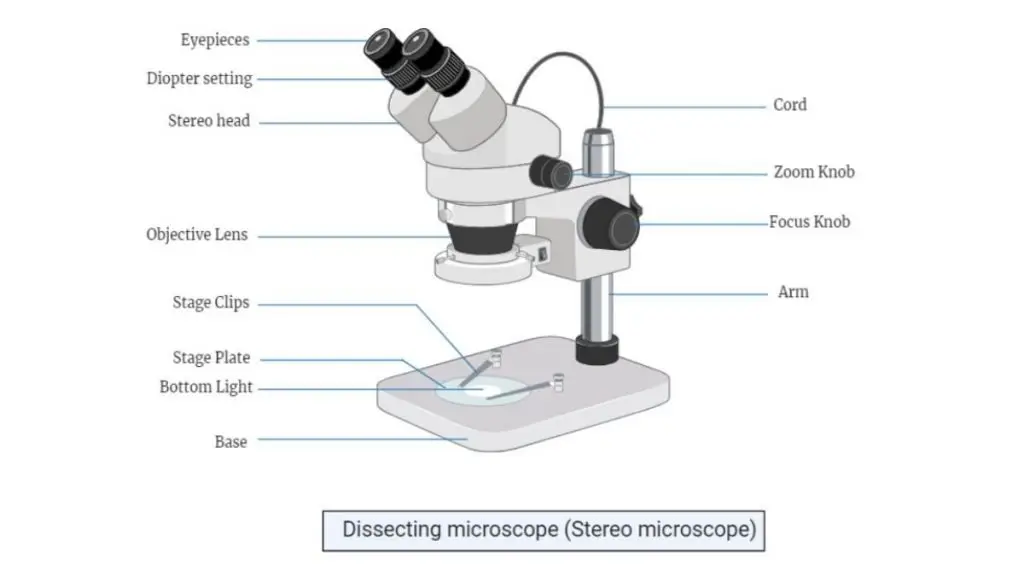
Dissecting Microscope Parts (Parts of Stereo microscope)
- LED Illuminators – The dissecting microscopes some have built-in lights such as a fixed LED illuminator.
- Eyepieces- They each have a different pathway of light into and out of the specimen. Each one has its own degree of magnification. And to get greater magnification, you can add some auxiliary eyepieces.
- Objective lenses: These also have different magnifications, focusing the image on the digital camera, and auxiliary objectives are employed to improve magnification.
- Stage- This is the instrument used to position the sample. They are big, so they can hold large specimen equipment.
- Optics System – anything between the fixed and zoom magnification that provides a fixed-focus lens.
- Digital camera – it is temporarily mounted on most dissecting microscopes for taking pictures, and it records the result of the pictures. This technology takes both 2D and 3D images (if you have the 3D digital camera).
Types of Dissecting microscopes (Stereo microscope)
Dissecting or stereo microscopes are for looking at 3D objects. These instruments represent different varieties for different uses, unique characteristics, unique magnification abilities. The next few entries describe the major types of dissection microscopes.
- Stereo Zoom Dissecting Microscope – This category has trinocular or binocular, zoom ration 6.7x to 45x. They could also have a digital camera to take images of the specimens they are viewing. A feature of these would be the double LED illuminator and the head that rotates 360°, which allows for extra viewing flexibility. You can adjust magnification by adding secondary objectives or eyepieces, which allows for more versatility for different jobs.
- Digital Tablet Dissecting Microscope – These microscopes are a higher level – a touch-screen LCD tablet camera with continuous magnification zoom from 6.7x up to 45x. Auxiliary eyepieces permit changes in magnification—these are dialed into more detail by switching on additional lenses on the objectives. The built-in 5.0-megapixel digital camera enables image and video capture that can be saved directly to the tablet or downloaded via a USB cable connection. They have integral LED illuminators at the head and foot that work separately to provide perfect lighting.
- Stereo Zoom Boom Stand Microscopes. – These have large bases and large platforms and can fit bigger specimens for examination. Their zooming range is from 6x to 45x, and can even go further by using auxiliary lenses or eyepieces. These microscopes use LED lights or dual pipe lighting so that adequate light strikes the specimen evenly.
- Stereo Zoom Dissecting Microscope–The small version has a zoom of 10x to 30x and a crisp, parfocalled view. The revolving head allows the eyepiece to be rotated either toward or away from the object, increasing its utility. It’s illuminated with a 10-watt halogen bulb and a 5-watt fluorescent bulb, providing adequate lighting to see distinctly.
- Dual Power Dissecting Microscope – With 10x and 30x magnifications, and a 360° rotating head, this microscope provides more observation flexibility. The second pair of objectives is parfocal, parcentered, and achromatic, so the image remains the same without any gray out. The lenses can be turned to change the amount of magnification. Also, an LED intensity light ring is used to illuminate the surface of the specimen evenly, and a flexible stand permits the viewing of larger objects at different heights.
- Single Power Stereo Dissection Microscope- They have low magnifications of about 10x to 40x. These have inclined eyepieces at 45° and diopter adjustments for 50mm-70mm. All of these qualities lend themselves well to a job that demands less detailed observation.
- Single Magnification Handheld Pocket Microscope- From Japan, portable microscope with two fixed magnification powers. There is no need for outside lighting, just straight-through high-quality optical glass. It is nice and small, so it is ideal for field work and just portable necessities in general.
Operating Procedure of Dissecting microscope
Like most microscopes that are used for examining larger specimens, the dissecting microscope (also known as stereoscopic microscope) can be utilised to study fossils, rocks, insects, and plant pieces, whilst they can also examine specimens mounted on slides.
The stages- These microscopes generally have two types of stages which can be used for viewing and dissecting:
- The first type can be a black or white opaque stage – This design is widely employed when observing non-transparent specimens
- Glass stage (transparent or opaque) – It can be used when illumination from below is required, particularly when the specimen is mounted on slides.
Installing the Stage- Before powering on the unit, the proper stage must be selected. When replacing an existing stage, these steps should be followed:
- Loosen the stage plate lock screw to remove the current stage
- When installing a glass stage, a blue filter is generally inserted into the stage’s central base
- The stage can be secured by tightening the locking screw
- Then reduce brightness to its lowest setting to protect the bulb’s life
Powering and Positioning- Next, activate the microscope using the on/off switch located on the base. The incident or transmitted light (or both depending on specimen) should then be activated. The body/head containing objectives must be lowered slowly using the coarse focus knob, which serves as a crucial foundation.
Eyepieces- The interpupillary distance needs adjustment by viewing through ocular lens and gently adjusting them inwards or outwards (similar to binoculars). Without a specimen present, a single circle should be visible through eyepieces after adjustment.
Specimen Mounting and focusing- The mounting process varies with specimen type:
- For specimens like Hydra that require mounting on glass slides, both incident and transmitted light can be used
- For opaque specimens (such as rocks), incident light is sufficient
After positioning the specimen in the focal plane, focusing can begin by gradually turning the focus knob until achieving sharp image clarity.
Zooming and Final steps- After initial focusing, the zoom knob can be used to examine specific areas with a lot of ease. When viewing structures like hydra tentacles, zooming may require additional focus adjustment as the image might become fuzzy.
The intensity controls allow for light emission adjustment and contrast modification.
At completion, these steps must be followed:
- Return zoom to minimum level
- Reduce light intensity
- Power off using on/off switch
- Cover with appropriate microscope cover/bag.
Applications of Dissecting microscope (Stereo microscope)
Like most microscopes that are utilised in various fields, the dissecting microscope (also known as stereoscopic microscope) has widespread applications across manufacturing, medical studies, quality control processes, inspection procedures and biomedical investigations.
These applications can be categorised into following areas-
- Manufacturing and Quality Control-
- Watch manufacturing processes that require high-powered magnification
- Circuit board assembly and detailed inspection procedures
- When quality checks need to be performed with a lot of ease.
- Medical and Surgical Applications- This microscope can be utilised for:
- Performing intricate microsurgical procedures where they need detailed visualization
- Conducting dissections that require precise magnification power
- Examining specimens under various zooming ranges.
- Forensic and Engineering Studies- The use of stereo microscope includes:
- Detailed inspection of fractures through fractography
- Forensic engineering investigations where it can provide crucial details
- Examining solid sample topography with enhanced clarity
- Biomedical and Research Applications –
- Entomological studies focusing on insect examination
- Detailed specimen analysis that requires stereoscopic viewing
- Generally employed when specimens need thorough inspection.
- Quality Assessment- They have significant applications in:
- Manufacturing sectors requiring precise measurements
- Industrial inspection processes where quality is paramount
- Examination procedures that need enhanced magnification.
- Inspection Procedures- The microscope finds extensive use in:
- Detailed component analysis
- Manufacturing defect identification
- Quality control processes that demand high precision viewing.
- Research and Development-
- Used extensively in biomedical research
- Employed for detailed specimen examination
- Applied in various scientific investigations
Advantages of Dissecting microscope
Like most microscopes that serve multiple purposes, the dissecting microscope (also known as stereoscopic microscope) possesses distinct advantages that make it one of the most significant microscopic techniques.
- Versatility in Applications- These microscopes can be utilised across various fields, which makes them exceptionally valuable when different types of specimens need examination with a lot of ease.
- Optical Advantages-
- The use of dual light pathways offers enhanced magnification power
- It can provide different zooming ranges for detailed visualization
- They have superior image clarity through stereoscopic viewing.
- Imaging capabilities- This microscope provides:
- Digital camera attachment options for recording specimens
- Enhanced documentation possibilities through imaging
- Superior recording capabilities that aid in research
- Practical Benefits – The stereoscopic microscope offers:
- Easy portability that allows movement between locations
- Simple operational procedures making it user-friendly
- Convenient handling during specimen examination.
- Specimen Viewing advantages –
- Generally allows examination of complete specimens
- It can view objects in their entirety rather than sections
- The use of whole-specimen viewing provides comprehensive analysis
- Operational Convenience- These microscopes provide:
- Straightforward operational procedures
- Enhanced ease of use during examination
- Simplified handling for various applications
- Technical Benefits-
- Superior magnification capabilities through dual pathways
- Enhanced image clarity for detailed examination
- Comprehensive viewing options for various specimens.
Disadvantages of Dissecting microscope
Like most microscopes that have certain limitations, the dissecting microscope (also known as stereoscopic microscope) presents specific disadvantages that affect its functionality and accessibility.
- Cost Implications-
- The Galilean Optical Systems, which form an essential component, can be quite expensive
- These microscopes generally require significant financial investment
- It can be costly to acquire and maintain these systems.
- Magnification Limitations- They have restricted capabilities including:
- Limited zooming range that typically doesn’t exceed 100x
- Reduced magnification power compared to other microscopes
- The use of lower powered objectives restricts detailed viewing.
- Structural Examination constraints- This microscope presents limitations when:
- Examining minute tissue structures that require high magnification
- Viewing specimens that need detailed cellular observation
- Generally analysing structures requiring powerful magnification.
- Technical Restrictions –
- Limited capability for high-powered examination
- Restricted usage for detailed cellular studies
- The use is confined to lower magnification observations
- System Limitations- These microscopes are restricted by:
- Complex optical systems that increase procurement costs
- Expensive components that affect accessibility
- Limited application in high-magnification studies.
- Practical Constraints-
- Unable to provide detailed cellular examination
- It can be restrictive for minute structure observation
- Generally unsuitable for high-magnification requirements.
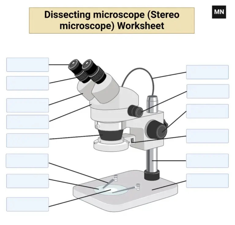
Dissecting Microscope Worksheet
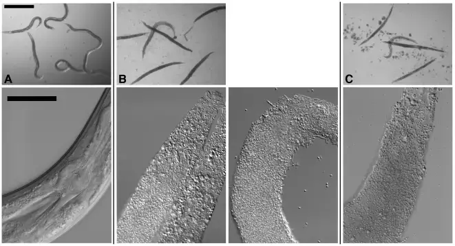
Dissecting microscope Sample images

- https://www.slideshare.net/waleedtareen2/stereo-microscope-or-dissecting-miscrscope-54430261
https://en.wikipedia.org/wiki/Stereo_microscope
https://www.microscopeinternational.com/what-is-a-stereo-microscope/
https://www.news-medical.net/life-sciences/What-are-Stereo-Microscopes-Used-For.aspx
https://www.microscope.com/education-center/five-things-you-should-know/about-stereo-microscopes
https://www.microscope-detective.com/stereo-microscope.html