What is transmission electron microscope?
Transmission Electron Microscope (TEM) – A TEM is a high-resolution microscope that uses a beam of electrons to pass through a thin sample, creating detailed images of the internal structure at the atomic or molecular level. It is used in material science, biology, and nanotechnology to examine ultrastructure.
This is one of the powerful electron microscopes that function on a beam of electrons that focuses on a specimen, thereby producing a highly magnified and detailed image of the specimen.
The power of magnification is much more than 2 million times better than that of the light microscope and produces the image of the specimen which in turn, allows the easy characterization of the image in its morphological features, compositions, and crystallization information as well.
Early discovery of cathode rays, like electrons, by Louis de Broglie in the early 1920s opened up a route to developing the electron microscope that utilized a beam of electrons to create a form of wave motion.
Magnetic fields worked as lenses for the electrons. From these inventions, the first electron microscope was invented by Ernst Ruska and Max Knolls in 1931 and transformed into a Transmission Electron Microscope (TEM) by Ernst Ruska with the help of Sieman’s company in 1933.
There are several advantages of this TEM microscope as compared to the light microscope whose efficiency is also very high.
Among all microscopes both light and electron microscopes, TEM are the most powerful microscopes used in laboratories. It can magnify a mall particle of about 2nm, and therefore they have a resolution limit of 0.2um.
Principle of Transmission Electron Microscope (TEM)
The TEM is based on the fundamental principle of substituting electrons instead of light for producing high-resolution images of specimens. In contrast to the light microscopes, which utilize visible light, the TEM makes use of a highly focused beam of electrons passing through an ultrathin specimen to produce an image in the process.
Electrons have much smaller wavelengths compared to light. For TEM, electrons’ wavelength is approximately 0.005 nm; that’s close to being 100,000 times shorter than that of light. A TEM can thereby provide a resolution around 1,000 times larger than what can be accomplished in light microscopes by detailed visualization of small structures.
If the incident beam of electrons traverses the specimen, it imparts images onto a fluorescent screen or a detector for ultrahigh-resolution images. This facilitates the observation of intricate internal structures like those present in viruses, organelles, or macromolecules with unparalleled clarity and precision. Thus, TEM is a very important tool in exploring the subcellular units and functions.
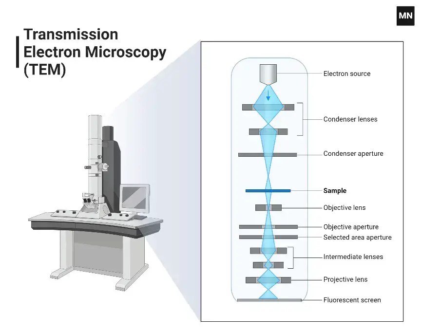
Parts of Transmission Electron Microscope (TEM)
A TEM consists of several important parts that have different functions in the imaging process.
- Electron Gun– The Electron Gun produces a beam of electrons that serves to illuminate the microscope.
- Vacuum System– The Vacuum System maintains a high vacuum environment inside the microscope so that the scattering of electrons by air molecules does not take place, thereby ensuring that the image taken will be clear and precise.
- Electron Lenses– Electron lenses use electromagnetic fields to focus and steer the electron beam onto the specimen, which then magnifies the transmitted electrons to produce a clear image.
- Specimen Stage– The specimen stage holds the ultra-thin specimen firmly in place, allowing for precise positioning and manipulation during the imaging process.
- Apertures– Apertures control the convergence of the electron beam and limit aberrations by selecting specific electron trajectories, thereby improving both contrast and resolution of the image.
- Detectors– Detectors capture electrons after they pass through the specimen, and an image is created. While old detectors are fluorescent screens and photographic film, new versions of transmission electron microscopes have electronic detectors that are charge-coupled devices (CCDs) or complementary metal-oxide-semiconductor cameras.
- Control Systems– Control systems include the computers and software dedicated to directing the TEM in the performance of its functions, including beam alignment, focusing, and image acquisition.
How does a Transmission Electron Microscope (TEM) work?
It is a very strong tool that allows scientists to see and study tiny samples at a really small level. Unlike the light microscopes, which mostly depend on visible light, TEMs use a stream of electrons to make very clear images. The way a TEM works can be divided into a few main steps.
- First, there is an electron gun at the top of the TEM that produces a stream of electrons. The most common form of electron gun uses a tungsten filament that is heated to very high temperatures. This heating causes electrons to be emitted from the filament surface through a process called thermionic emission.
- Then the emitted electrons are passed through several electromagnetic lenses. These are condenser lenses that conduct the electron beam to where it strikes the specimen and also control how wide and strong the beam is. The current in the condenser lenses can be manipulated by the operator to enhance the beam for a particular sample and the imaging conditions they desire.
- A very focused electron beam then strikes the sample, that is prepared with great care and placed on a small metal grid. As the electrons pass through the sample, a few get scattered by the atoms in the material. How much scattering takes place depends upon several factors, such as thickness of the sample, its density, and atomic number. Regions that are thicker or contain elements with higher atomic numbers will scatter electrons more strongly.
- Below the specimen, there are more electromagnetic lenses, which include the objective lenses and projector lenses. These lenses focus and make the transmitted electron beam larger. The objective lenses produce the first enlarged image of the specimen, and then the projector lenses enlarges that image to be used on the screen or a camera at the bottom of the TEM column. The magnification power of TEMs is pretty high, with advanced instruments able to enlarge images over one million times.
- The entire path of the electrons from the gun to the camera must be in a very high vacuum for the TEM to work properly. A small amount of air can scatter the electrons and thus make the image worse. Thus, the column of the microscope is pumped up to vacuum levels that are only comparable to outer space.
- Finally, the large electron beam strikes a fluorescent viewing screen, making it possible for the scientists to observe and record an image. Those areas of the specimen that scatter more electrons, or are thicker or denser, appear darker, while areas that allow more electrons to pass through are brighter. This makes the images seen under TEM typically black and white in contrast.
Methods used for Sample preparation for visualization by TEM
Sample preparation is the most important step in TEM for getting good-quality images and data. The method of preparation is dependent on the type of sample and the kind of information to be extracted. Some common techniques for TEM sample preparation are:
- Fixation: Crosslinks proteins and lipids of biological specimens with chemicals like glutaraldehyde and osmium tetroxide, thereby stabilizing them.
- Dehydration: Removes the water from biological specimens using a series of ethanol or acetone solutions so that there is no structural disruption when the images will be taken.
- Embedding: Infuses dehydrated samples in resins such as epoxy or acrylic, which helps support ultrathin sectioning.
- Sectioning: The ultrathin sections (50–70 nm) are prepared using an ultramicrotome with a diamond or glass knife by slicing embedded samples.
- Staining: Uses heavy metals like uranyl acetate or lead citrate, which bind to parts of cells to increase contrast.
- Cryofixation: Freezes samples quickly so their original state is preserved; typically used for delicate biological specimens.
- Ion Milling: A focused ion beam makes material samples thinner so that electrons can pass through without causing stress.
- Drop Casting: Deposes a diluted suspension of nanoparticles or fine powders onto the TEM grid and then allows evaporation of the solvent, which leaves a thin film suitable for imaging.
Step by step procedure;
Preparing samples for Transmission Electron Microscopy (TEM) is very important. It includes careful steps to make sure the samples are thin enough for electrons to pass through and do not have any unwanted marks. Here are the main steps for preparing samples for TEM viewing:
Sample Preparation Steps
- First Cutting/Sectioning- Start by cutting the bulk material into smaller pieces, which would be less than 100 µm thick. This can be done using various techniques, like a slow-speed diamond saw, ultrasonic cutter, or electro-discharge cutting. The aim here is to obtain a sample thickness of about 150-300 µm before further thinning.
- Thinning the Sample- Grind and polish the sample to approximately 5-15 µm. This is a preparation step that exposes the sample for further processes on a relatively scratch-free surface2. During grinding, apply gentle pressure to avoid creating cracks or damage in the specimen.
- Punching Discs- When the sheet is thin enough, punch out discs 3 mm wide. Be careful not to damage the edges since usually only the center part of the disc is used for looking at it.
- Dimpling- Dimple grinds the punched disc to make an area 2 to 10 microns thick on some parts of the disc, leaving a shallow dimple or pit in its center, permitting electrons to move through. End
- Final Polishing- For TEM imaging, final electron transparency is achieved using methods like jet polishing or ion beam milling. Ion beam milling is very effective since it removes material at a slow rate and does not damage the surface; therefore, this is a good surface to observe. Electrochemical etching can be used for conducting samples sometimes.
- Mounting on Grids- Mount the electron-transparent discs onto TEM grids once prepared. These grids are generally made of materials like copper, and in most cases, are coated with something similar to carbon to improve conductivity and prevent charging during imaging
- Cleaning and Coating- Clean the sample by removing residues that have been deposited by previous preparation steps. This is often done using solvents like chloroform or ethanol.
Optionally: carbon coat a thin layer on the surface of the sample to prevent any kind of glow discharge during TEM imaging.
Applications of Transmission Electron Microscope (TEM)
- Materials Science – TEM is used to look at the tiny structure, crystal form, and flaws in metals, ceramics, and polymers, helping to create better materials.
- Nanotechnology – TEM helps to analyze nanoparticles, nanowires, and other small structures, giving us information about their shape and makeup.
- Life Sciences – TEM is used to study the ultrastructure of cells, tissues, and viruses to better understand biological processes and mechanisms of disease.
- Semiconductor Research – TEM studies the microstructure of semiconductor devices, which determines defects and the quality of fabrication, very important to the electronics industry.
- Pharmaceuticals – TEM helps in characterizing drug compounds and delivery systems to ensure efficacy and safety in drug development.
- Geology– TEM studies the small structures of minerals and rocks, and helps us understand how geological processes work and the history of how Earth was formed.
- Forensic Analysis- TEM helps in examining forensic samples by studying small evidence under a microscope, which supports criminal investigations.
Advantages of Transmission Electron Microscope (TEM)
- Very high resolution. Images from the TEM are quite sharp, showing atomic-level structure.
Powerful magnification: Structures can be enlarged to a view exceeding one million times, resulting in detailed images of tiny features. - It is easy to study various features of the sample with the help of TEM by using different imaging modes, including dark/bright field and phase contrast.
- Electron diffraction patterns from small areas can be obtained using TEM to give information about the crystallographic structure of materials.
- High-quality images are produced by TEM that helps in detailed analysis.
- Permanent Image Capture, TEM captures permanent images. These can be used as a record as well as further analysis.
Limitations of Transmission Electron Microscope (TEM)
- Sample Preparation- The TEM needs samples to be very thin, usually less than 100 nanometers, to allow electrons to pass through. This can be time-consuming and can change the natural state of the sample.
- Vacuum Environment- Samples must be placed in a high-vacuum environment so that air molecules do not scatter the electrons. This makes TEM unsuitable for observing living specimens.
- Radiation Damage- The exposure to the electron beam holds the potential to harm delicate samples, including biological specimens and polymers, which could result in structural alterations during the imaging process.
- Limited Field of View- The Transmission Electron Microscope offers a comparatively small field of view, which might not accurately reflect the entirety of the sample, thereby possibly introducing sampling bias.
- Complex Image Interpretation- TEM images represent two-dimensional projections of three-dimensional structures, meaning that complex analysis and interpretation is required to make an accurate morphological understanding of the sample.
- Instrumental Constraints- TEM instruments are large and expensive, which requires specialized facilities and maintenance and may limit the accessibility of TEM for some researchers.
- https://www.news-medical.net/life-sciences/Sample-Preparation-in-TEM.aspx
- https://warwick.ac.uk/fac/sci/physics/current/postgraduate/regs/mpagswarwick/
- https://www.jeol.co.jp/en/science/em.html
- https://www.britannica.com/technology/electron-microscope
- https://www.ccber.ucsb.edu/collections-botanical-collections-plant-anatomy/transmission-electron-microscope
- https://www.news-medical.net/life-sciences/What-is-Transmission-Electron-Microscopy.aspx
- https://www.microscopemaster.com/transmission-electron-microscope.html
- https://en.wikipedia.org/wiki/Transmission_electron_microscopy
- https://www.slideshare.net/TakeenKhurshid/transmission-electron